When there’s a clear, documented way to diagnose vertebrogenic pain.
That’s Living Proof.

The Background
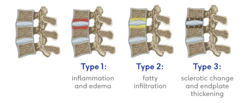
To confirm that a patient has vertebrogenic pain, physicians use MRI to look for specific changes that occur with endplate inflammation, which are called Modic changes. They’re called “Modic changes” because in 1988, Dr. Michael Modic was the first to publish on identifying and classifying degenerative endplate and marrow changes surrounding a dehydrated intervertebral disc. There were three types of bone marrow changes identified: Types 1, 2, and 3. Types 1 and 2 are the ones that can be used to identify vertebrogenic pain.
Modic Type 1
Normal bone contains trabeculae, or internal scaffolding. In the spaces between the trabeculae there is red bone marrow, which produces
blood cells. In Modic Type 1 changes, you see:
- Vascular development in the vertebral body
- Findings of inflammation and edema
- NO trabecular damage or marrow changes

Modic Type 2
Normal bone contains trabeculae, or internal scaffolding. In the spaces
between the trabeculae there is red bone marrow, which produces
blood cells. In Modic Type 2 changes, you see:
- Changes in bone marrow
- Fatty replacement of formally red cellular marrow
- Marrow is substituted with visceral fat
- Same fat located on hips and bellies

Intracept Procedure Indications
- Chronic low back pain of at least 6 months duration; and
- Failure to respond to at least 6 months of conservative care; and
- MRI demonstrated Modic Type 1 or Type 2 changes at one or more vertebrae from L3 to S1 documented by at least one of the following:
- Modic Type 1 and/or Modic Type 2
–Endplate changes, inflammation, edema, disruption, and/or fissuring
–Fibrovascular bone marrow changes (hypointensive signal for Modic Type 1)
–Fatty bone marrow replacement (hyperintensive signal for Modic Type 2)
Let’s Get to It:
What the Intracept Procedure Involves
The Intracept Procedure is a minimally invasive, outpatient procedure for patients with vertebrogenic pain. The procedure targets a specic nerve within the vertebra called the basivertebral nerve and has been shown to improve function and relieve pain long-term. The procedure is implant-free, preserving future treatment options for other spine
conditions. Here’s what the procedure involves.
Step One: Access the Pedicle
Under fluoroscopic guidance, the Intracept Introducer Cannula Assembly is advanced through the pedicle.
Step Two: Create the Channel
The Intracept Curved Cannula Assembly is used to create a channel to the trunk of the basivertebral nerve.


Step Three: Place the RF Probe
The Intracept RF Probe is inserted into the curved path and placed at the trunk of the basivertebral nerve.
Step Four: Ablate the BVN
The Intracept RF Generator is used to deliver radiofrequency energy that ablates the basivertebral nerve.

Why Choose VIP SPECIALISTS for MILD Procedure?
Our team of experienced physicians specializes in providing non-surgical options for spine pain. We are committed to patient well-being and use the latest techniques and technology to deliver the highest standard of care. If you’re seeking a non-surgical solution for spine pain, contact us today for a consultation.

